Our research is focused on the development and use of novel sophisticated electron-optical instruments and devices for multichannel spin filtering.
SPECTRAL RANGES AND FACILITIES
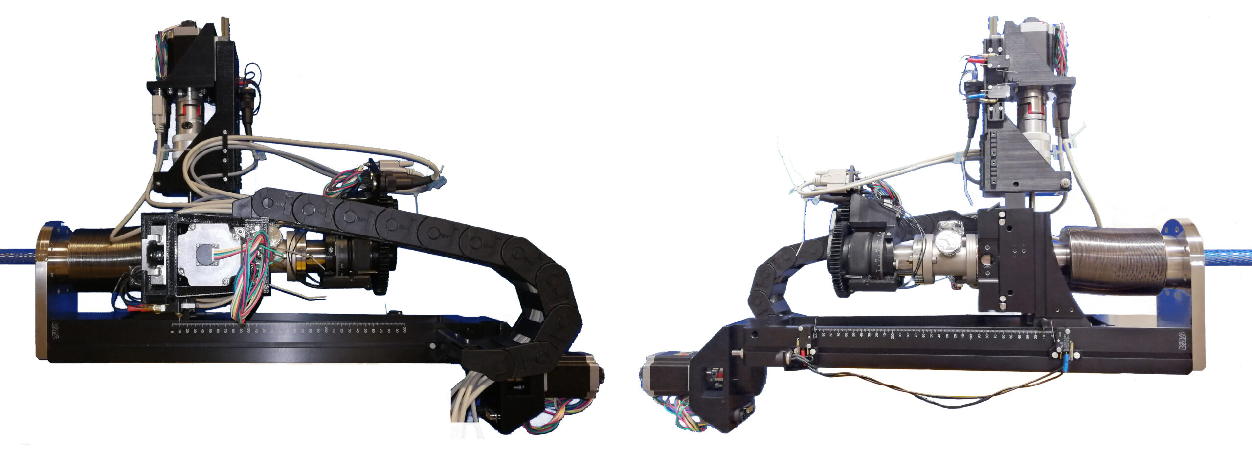
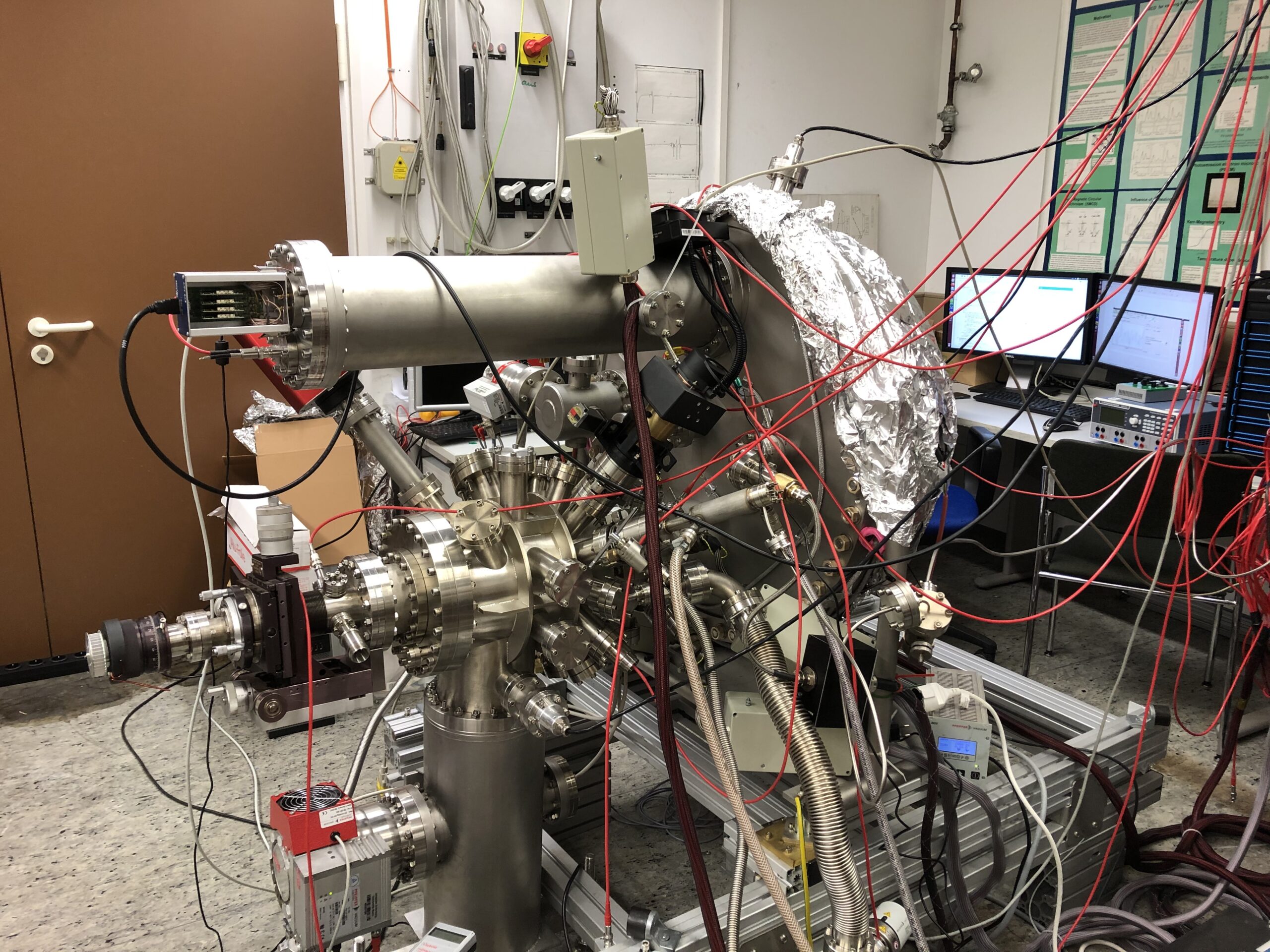
Among the key instrumentation, we developed various kinds of time-of-flight momentum microscopes [1]. The low-energy microscope has been optimized for high-resolution band mapping with a k-field-of-view up to a diameter of 7 Å-1 and kinetic energies up to 1.5 keV. A modified high-energy optics targets efficient electron detection in the HAXPES range with kinetic energies up to > 7.5 keV and large k-fields-of-view up to > 22 Å-1, as needed for hard X-ray photoelectron diffraction (hXPD).

A second technological stronghold is the development of imaging spin filters [2]. First experiments have been performed using 2D-recording with hemispherical analyzers, either in the conventional (E-θ) detection mode in our lab or as momentum microscope with (kx-ky) recording at the Max-Planck Institute in Halle. Later, we established 3D-recording (kx-ky-τ) using the time-of-flight τ for encoding the energy coordinate. On the quest for the optimum spin filter the Ir(001)-surface with pseudomorphic Au-monolayer has proven “ideal”, especially due to life times of months (work in Halle). For 3D recording including ToF a spin filter with larger usable energy range would be desirable. For work in the shielding hutches at hard-X-ray beamlines and FELs, a fully remote-controlled version of imaging spin filter has been developed.
Experiments covered spectral ranges from < 6eV to > 7 keV, in particular: near-threshold emission using lab sources [3], the vacuum ultraviolet range at BESSY II [4], soft X-rays at PETRA III, beamline P04 (and in the future at DIAMOND, UK) to hard X-rays at PETRA III, beamline P22 [5]. The instrumental key feature is an increase of the dimensionality of the recording scheme from 2D to 3D.
SCIENTIFIC CASES
Besides detailed mapping of electronic bands in a number of metallic and semiconducting systems with an emphasis on anomalous states [6] and organic charge-transfer materials [7] the method has been used for the study of plasmonic electron emission [8]. A running development is the combination of multichannel spin-filtering with bulk-sensitive photoemission in the soft and hard X-ray range [9]. At the end of a decade-long development with a lot of experience gained from early work at SPring 8 in Japan, the first spin-resolved HAXPES experiments became feasible at PETRA III, beamline P22, after implementation of 3D parallel spin recording. Another upcoming important application is the use of time-resolved recording [10]. Our alternative highly-effective approach to hard-X-ray photoelectron spectroscopy (ToF-HAXPES) [11] constitutes a novel tool for full-field imaging X-ray photoelectron diffraction (XPD) [12] which has led to a new understanding of the various contributions of Laue- and Kikuchi-type XPD [13].
CURRENT DEVELOPMENTS
Among the running technical developments, the understanding and reduction of the space-charge effect in short-pulse excitation [14] is a central issue. The method was extended towards high-resolution electron energy-loss microscopy using an ultra-cold atom photoemission source in cooperation with groups in Orsay [15]. The search for an optimum spin filter for 3D ToF recording is still going on and spin-filter systems with exchange-scattering asymmetry are explored. The most recent development is a combination of dispersive energy filtering based on a single hemispherical analyzer with time-of-flight recording, as a booster for ultimate resolution [16]. k-resolved detection circumvents the problem of different transit times of electrons traveling on different ray paths through a hemispherical analyzer [17].
USE OF THE ToF k-MICROSCOPE AT FREE-ELECTRON LASERS AND HHG-SOURCES
Angle-resolved photoelectron spectroscopy with time resolution (TR-ARPES) down to the fs range is a challenging task due to the inherent problems to record electrons with high charge density in a sub-ps instance (Coulomb interaction, particle optics in general). Despite these serious obstacles (in comparison with photon recording), there are strong efforts worldwide to establish TR-ARPES due to the unique information content in fully k-resolved photoemission data. Thanks to its capability of multi-dimensional recording, the time of flight k-microscope seems a very promising alternative to the conventional use of hemispherical analyzers with their inherent restriction of the simultaneously recorded phase-space volume. Next to many decades of development of hemispherical analyzers, reaching impressive results in energy resolution down to the principal limits, ToF k-microscopy is still in its infancy. Nevertheless, first pilot experiments have been performed using the time-of-flight momentum microscope by a few pioneering groups at the free-electron laser FLASH [18] and several HHG-sources [19]. A big task in this context is the development of suitable data harvesting architectures for multidimensional recording [20].
References:
[1] [back] Development of time-of-flight photoelectron momentum microscopes:
ToF k-microscopes for low energies
1.1 Direct 3D mapping of the Fermi surface and Fermi velocity. K. Medjanik, O. Fedchenko, S. Chernov, D. Kutnyakhov, M. Ellguth, A. Oelsner, B. Schönhense, T.R.F. Peixoto, P. Lutz, C.-H. Min, F. Reinert, S. Däster, Y. Acremann, J. Viefhaus, W. Wurth, H.-J. Elmers and G. Schönhense,
Nature Mat. 16, 615 (2017)
1.2 Space-, Time- and Spin-resolved Photoemission; G. Schönhense, K. Medjanik and H.-J. Elmers
Journal of Electron Spectrosc. Relat. Phenom. 200, 94 (2015) (review)
ToF k-microscope for high energies
1.3 Progress in HAXPES Performance Combining Full-Field k-Imaging with Time-of-Flight Recording; K. Medjanik, S. V. Babenkov, S. Chernov, D. Vasilyev, B. Schönhense, C. Schlueter, A. Gloskowskii, Yu. Matveyev, W. Drube, H. J. Elmers and G. Schönhense
J. of Synchr. Radiation 26, 1996–2012 (2019)
[2] [back] Development of imaging spin filters:
Imaging spin filter in combination with conventional hemispherical analyzers
2.1 Highly Efficient Multichannel Spin-Polarization Detection; M. Kolbe, P. Lushchyk, B. Petereit, H.-J. Elmers, G. Schönhense, A. Oelsner, C. Tusche and J. Kirschner
Phys. Rev. Lett. 107, 207601 (2011)
2.2 Vectorial spin-polarization detection in multichannel spin-resolved photoemission spectroscopy using an Ir(001) imaging spin filter; E.D. Schäfer, S. Borek, J. Braun, J. Minar, H. Ebert, K. Medjanik, D. Kutnyakhov, G. Schönhense and H.-J. Elmers
Phys. Rev. B 95, 104423 (2017)
2.3 Specular reflection of spin-polarized electrons from a W(001) spin-filter crystal in a larger range of scattering energies and angles, D. Kutnyakhov, H.J. Elmers, G. Schönhense, C. Tusche, S. Borek, J. Braun, J. Minàr and H. Ebert
Phys. Rev. B 91, 014416 (2015)
2.4 Direct observation of half-metallicity in the Heusler compound Co2MnSi, M. Jourdan, J. Minár, J. Braun, A. Kronenberg, S. Chadov, B. Balke, A. Gloskovskii, M. Kolbe, H.J. Elmers, G. Schönhense, H. Ebert, C. Felser and M. Kläui
Nature Commun. 5, 3974(2014)
2.5 Imaging Spin Filter for Electrons Based on Specular Reflection from Ir (001), D. Kutnyakhov, P. Lushchyk, D. Perriard, M. Kolbe, K. Medjanik, E. Fedchenko, S.A. Nepijko, H.J. Elmers, G. Salvatella, R. Gort, T. Bähler,T. Michlmayer, A. Fognini, Y. Acremann, A. Vaterlaus, C. Tusche, A. Krasyuk, J. Kirschner, F. Giebels, H. Gollisch, R. Feder and G. Schönhense
Ultramicroscopy 130, 63 (2013)
2.6 Tabletop-based setup for spin-, time- and angle-resolved photoemission spectroscopy, K. Bühlmann, R. Gort, A. Fognini, S. Däster, S. Holenstein, N. Hartmann, Y. Zemp, G. Salvatella,T. U. Michlmayr, T. Bähler, D. Kutnyakhov, K. Medjanik, G. Schönhense, A. Vaterlaus and Y. Acremann,
submitted
Rev. Sci. Instrum. 91, 063001 (2020)
Imaging spin filter in combination with a momentum microscope with twin-hemispherical analyzer
2.7 Spin resolved photoelectron microscopy using a two-dimensional spin-polarizing electron mirror; C. Tusche, M. Ellguth, A. A. Ünal, C.-T. Chiang, A. Winkelmann, A. Krasyuk, M. Hahn, G. Schönhense and J. Kirschner
Appl. Phys. Lett. 99, 032505 (2011)
2.8 Efficient Spin-Polarization Analysis in Photoelectron Emission Microscopy With an Imaging Spin Filter, C. Tusche, M. Ellguth, A. Krasyuk, A. Winkelmann, D. Kutnyakhov, P. Lushchyk, K. Medjanik, G. Schönhense and J. Kirschner
Ultramicroscopy 130, 70 (2013)
Imaging spin filter in combination with a time-of-flight momentum microscope
2.9 Spin-Filtered Time-of-Flight k-Space Microscopy of Ir – Towards the “Complete” Photoemission Experiment; G. Schönhense, K. Medjanik, S. Chernov, D. Kutnyakhov, O. Fedchenko, M. Ellguth, D. Vasilyev, A. Zaporozhchenko, D. Panzer, A. Oelsner, C. Tusche, B. Schönhense, J. Braun, J. Minár, H. Ebert, J. Viefhaus, W. Wurth and H. J. Elmers
Ultramicroscopy 183, 19–29 (2017)
[3] [back] Near-threshold photoemission using 6 eV photons:
3.1 Band structure tuning of Heusler compounds revisited: Spin- and momentum-resolved electronic structure analysis of compounds with different band filling; S. Chernov, C. Lidig, O. Fedchenko, K. Medjanik, S. Babenkov, D. Vasilyev, M. Jourdan, G. Schönhense and H. J. Elmers
arXiv 1910.05205 (2019) and submitted
3.2 Energy- and k-resolved mapping of the magnetic circular dichroism in threshold photoemission from Co films on Pt(111); M. Staab, D. Kutnyakhov, R. Wallauer, S. Chernov, K. Medjanik, H.- J. Elmers, M. Kläui and G. Schönhense
Phys. Rev. B 95, 165437 (2017)
[4] [back] VUV photoemission at BESSY II:
4.1 Multi-MHz Time-of-Flight Electronic Bandstructure Imaging of Graphene on Ir(111); C. Tusche, P. Goslawski, D. Kutnyakhov, M. Ellguth, K. Medjanik, H. J. Elmers, S. Chernov, R. Wallauer, D. Engel, A. Jankowiak, and G. Schönhense
Appl. Phys. Lett. 108, 261602 (2016)
4.2 Spin texture of time-reversal symmetry invariant surface states on W(110); D. Kutnyakhov, S. Chernov, K. Medjanik, R. Wallauer, C. Tusche, M. Ellguth, S.A. Nepijko, M. Krivenkov, J. Braun, S. Borek, J. Minar, H. Ebert, H.-J. Elmers and G. Schönhense
Scientific. Rep. 6, 29394 (2016)
[5] [back] Soft- and hard-X-ray photoemission at PETRA III:
5.1 Direct 3D mapping of the Fermi surface and Fermi velocity. K. Medjanik, O. Fedchenko, S. Chernov, D. Kutnyakhov, M. Ellguth, A. Oelsner, B. Schönhense, T.R.F. Peixoto, P. Lutz, C.-H. Min, F. Reinert, S. Däster, Y. Acremann, J. Viefhaus, W. Wurth, H.-J. Elmers and G. Schönhense,
Nature Mat. 16, 615 (2017)
5.2 Relation between Spin-Orbit Induced Spin Polarization, Fano-Effect and Circular Dichroism in Soft X-ray Photoemission; D. Vasilyev, K. Medjanik, S. Babenkov, M. Ellguth, G. Schön-hense and H.-J. Elmers,
J. Phys. C 32, 135501 (2020)
5.3 4D texture of circular dichroism in soft-x-ray photoemission from tungsten; O. Fedchenko, K. Medjanik, S. Chernov, D. Kutnyakhov, M. Ellguth, A. Oelsner, B. Schönhense, T. Peixoto, P. Lutz, C.-H. Min, F. Reinert, S. Däster, Y. Acremann, J. Viefhaus, W. Wurth, J. Braun, J. Minár, H. Ebert, H. J. Elmers and G. Schönhense
New J. of Phys. 21, 013017 (2019)
5.4 Progress in HAXPES Performance Combining Full-Field k-Imaging with Time-of-Flight Recording; K. Medjanik, S. V. Babenkov, S. Chernov, D. Vasilyev, B. Schönhense, C. Schlueter, A. Gloskowskii, Yu. Matveyev, W. Drube, H. J. Elmers and G. Schönhense
J. of Synchr. Radiation 26, 1996–2012 (2019)
5.5 Néel vector induced manipulation of electronic states in the collinear antiferromagnet Mn2Au; H. J. Elmers, S. V. Chernov, S. P. Bommanaboyena, S. Yu. Bodnar, K. Medjanik, S. Babenkov, O. Fedchenko, D. Vasilyev, S. Y. Agustsson, C. Schlueter, A. Gloskovskii, Yu. Matveyev, Y. Skourski, J. Minar, L. Smejkal, J. Sinova, M. Kläui, G. Schönhense and M. Jourdan
submitted (2020)
[6] [back] Surface states:
6.1 Hosting of surface states in spin-orbit induced projected bulk band gaps of W(110) and Ir(111); H. J. Elmers, D. Kutnyakhov, S.V. Chernov, K. Medjanik, O. Fedchenko, A. Zaporozhchenko-Zymakova, M. Ellguth, C. Tusche, J. Viefhaus, G. Schönhense
J. Phys.: Condens. Matter 29 (2017) 255001
6.2 Rashba splitting of the Tamm surface state on Re(0001) observed by spin-resolved photo-emission and scanning tunnelling spectroscopy; H.J. Elmers, J. Regel, T. Mashof, J. Braun, S. Babenkov, S. Chernov, O. Fedchenko, K. Medjanik, D. Vasilyev, J. Minar, H. Ebert and G. Schönhense,
Phys. Rev. Research 2, 013296 (2020)
6.3 Anomalous d-like Surface Resonance on Mo(110) Analyzed by Time-of-Flight Momentum Microscopy; S. Chernov, K. Medjanik, D. Kutnyakhov, C. Tusche, S. A. Nepijko, A. Oelsner, J. Braun, J. Minár, S. Borek, H. Ebert, H. J. Elmers, J. Kirschner and G. Schönhense
Ultramicroscopy 159, 463 (2015)
6.4 Spin mapping of surface and bulk Rashba states in ferroelectric α-GeTe(111) films; H. J. Elmers, R. Wallauer, M. Liebmann, J. Kellner, M. Morgenstern, R. N. Wang, J. E. Boschker, R. Calarco, J. Sanchez-Barriga, O. Rader, D. Kutnyakhov, S. V. Chernov, K. Medjanik, C. Tusche, M. Ellguth, H. Volfova, S. Borek, J. Braun, J. Minár, H. Ebert and G. Schönhense
Phys. Rev. B 94, 201403 (2016)
6.5 Momentum-resolved photoelectron absorption in surface barrier scattering on Ir(111) and graphene/Ir(111); A. Zaporozhchenko-Zymaková, D. Kutnyakhov, K. Medjanik, C. Tusche, O. Fedchenko, S. Chernov, M. Ellguth, S.A. Nepijko, H.J. Elmers and G. Schönhense
Phys. Rev. B 96, 155108 (2017)
[7] [back] Organic charge-transfer systems:
7.1 Electron- and X-Ray Spectroscopies of Organic Charge-Transfer Complexes; K. Medjanik, H.-J. Elmers, G. Schönhense, J.-P.l Pouget, R. Valenti and M. Lang
Phys. Status Solidi B 1800745 (2019), (review article)
7.2 Orbital-Resolved Partial Charge Transfer from the Methoxy Groups of Substituted Pyrenes in Complexes with Tetracyanoquonodimethane – a NEXAFS Study; K. Medjanik, D. Chercka, P. Nagel, M. Merz, S. Schuppler, M. Baumgarten, K. Müllen, S.A. Nepijko, H.-J. Elmers, G. Schönhense, H.O. Jeschke and R. Valenti
J. Am. Chem. Soc. 134, 4694 (2012)
[8] [back] Plasmonic electron emission:
8.1 Momentum distribution of electrons emitted from resonantly excited individual gold nanorods; M. Lehr, B. Foerster, M. Schmitt, K. Krüger, C. Sönnichsen, G. Schönhense and H.-J. Elmers,
Nano Lett. 17, 6606 (2017)
8.2 Evidence of Spatially Inhomogeneous Electron Temperature in a Resonantly-Excited Array of Bow-Tie Nanoantennas; M. Lehr, K. Bley, N. Vogel, B. Rethfeld, G. Schönhense and H.-J. Elmers,
J. Phys. Chem. C 12, 319, 12429-12436 (2019)
8.3 Polarization dependence of plasmonic near-field enhanced photoemission from cross antennas; P. Klaer, G. Razinskas, M. Lehr, X.-F. Wu, B. Hecht, F. Schertz, H.-J. Butt, G. Schönhense and H.-J. Elmers,
Appl. Phys. B 122, 136 (2016)
8.4 Near Field of Strongly Coupled Plasmons: Uncovering Dark Modes; F. Schertz, M. Schmelzeisen, R. Mohammadi, M. Kreiter, H.-J. Elmers and G. Schönhense
Nano Lett. 12, 1885 (2012)
8.5 Field Emission of Electrons Generated by the Near Field of Strongly Coupled Plasmons; F. Schertz, M. Schmelzeisen, M. Kreiter, H.-J. Elmers and G. Schönhense
Phys. Rev. Lett. 108, 237602 (2012)
[9] [back] Spin-resolved bulk photoemission:
9.1 Relation between Spin-Orbit Induced Spin Polarization, Fano-Effect and Circular Dichroism in Soft X-ray Photoemission; D. Vasilyev, K. Medjanik, S. Babenkov, M. Ellguth, G. Schönhense and H.-J. Elmers,
J. Phys. C 32, 135501 (2020)
9.2 Spin-Resolved Bulk Electronic Structure Analysis of the Half-Metallic Heusler Ferromagnet Co2MnSi; S. Chernov, S. Babenkov, D. Vasilyev, K. Medjanik, O. Fedchenko, M. Jourdan, S. Andrieu, C. Guillemard, F. Bertran, P. LeFevre, M. Schmitt, C. Schlueter, Yu. Matveyev, A. Gloskowski, R. Claessen, H.-J. Elmers and G. Schönhense,
in preparation
9.3 Determination of the bulk spin polarization of Fe3O4 (111) thin films by means of spin-resolved hard X-ray time-of-flight microscopy; M. Schmitt, O. Kirilmaz, S. Chernov, S. Babenkov, D. Vasilyev, K. Medjanik, O. Fedchenko, Y. Matveyev, A. Gloskowski, C. Schlueter, H.-J. Elmers, G. Schönhense, M. Sing and R. Claessen,
in preparation
9.4 Development of hard X-ray photoelectron SPLEED-based spectrometer applicable for probing of buried magnetic layer valence states; X. Kozina, E. Ikenaga, C. E. Viol Barbosa, S. Ouardi, J. Karel, M. Yamamoto, K. Kobayashi, H.-J. Elmers, G. Schönhense and C. Felser
Journal of Electron Spectrosc. Relat. Phenom. 211, 12-18 (2016)
9.5 Spin Polarimetry and Magnetic Dichroism on a buried magnetic layer using Hard X-ray Photoelectron Spectroscopy; G. Stryganyuk, X. Kozina, G. H. Fecher, S. Ouardi, S. Chadov, C. Felser G. Schoenhense, P. Lushchyk, A. Oelsner, P. Bernhard, E. Ikenaga, T. Sugiyama, H. Sukegawa, Z. Wen, K. Inomata and K. Kobayashi
Japanese J. of Appl. Phys. 51 (2012) 016602
9.6 Magnetic dichroism in angle-resolved hard x-ray photoemission from buried layers; X. Kozina, G. H. Fecher, G. Stryganyuk, S. Ouardi, B. Balke, C. Felser, G. Schönhense, E. Ikenaga, T. Sugiyama, N. Kawamura, M. Suzuki, T. Taira, T. Uemura, M. Yamamoto, H. Sukegawa, W. Wang, K. Inomata and K. Kobayashi
Phys. Rev. B 84 (2011) 054449
[10] [back] Time-resolved photoemission:
10.1 Time and momentum-resolved photoemission studies using time-of-flight momentum microscopy at a free electron laser; D. Kutnyakhov, P. Xian, M. Heber, F. Pressacco, G. Mercurio, A. Benz, G. Wenthaus, H. Meyer, S. Gieschen, K. Bühlman, S. Däster, R. Gort, D. Curcio, K. Volckaert, M. Bianchi, Ch. Sanders, J. Miwa, S. Ulstrup, A. Oelsner, C. Tusche, Y.J. Chen, S.Y. Agustsson, D. Vasilyev, K. Medjanik, G. Brenner, S. Dziarzhytski, H. Redlin, J. Hauer, M. Dendzik, S. Dong, L. Rettig, J. Demsar, H.J. Elmers, Ph. Hofmann, R. Ernstorfer, G. Schönhense, Y. Acremann and W. Wurth
Rev. Sci. Instrum. 91, 013109 (2020)
[11] [back] Bulk bandmapping using hard X-rays:
11.1 High-accuracy bulk electronic bandmapping with eliminated diffraction effects using hard X-ray photoelectron momentum microscopy; S. Babenkov, K. Medjanik, D. Vasilyev, S. Chernov, C. Schlueter, A. Gloskovskii, Yu. Matveyev, W. Drube, B. Schönhense, K. Rossnagel, H.-J. Elmers and G. Schönhense
Comms. Phys. 2, 107 (2019)
11.2 Hard X-ray Photoelectron Momentum Microscopy and Kikuchi-Diffraction on InGaMnAs Thin Films; K. Medjanik, O. Fedchenko, O. Yastrubchak, J. Sadowski, L. Gluba, D. Vasilyev, S. Babenkov, S. Chernov, A. Winkelmann, H. J. Elmers and G. Schönhense,
Submitted, e-print on arXiv 2010.12359 (2020)
[12] [back] Hard X-ray photoelectron diffraction (hXPD):
12.1 High-resolution hard-X-ray Photoelectron Diffraction in a Momentum Microscope - the Model Case of Graphite; O. Fedchenko, A. Winkelmann, K. Medjanik, S. Babenkov, D. Vasilyev, S. Chernov, C. Schlueter, A. Gloskovskii, Yu. Matveyev, W. Drube, B. Schönhense, H. J. Elmers and G. Schönhense
New J. of Phys. 21, 113031 (2019)
12.2 Emitter-Site Specificity of Hard X-ray Photoelectron Kikuchi-Diffraction; O. Fedchenko, A. Winkelmann, S. Chernov, K. Medjanik, S. Babenkov, S. Y. Agustsson, D. Vasilyev, M. Hoesch, H.J. Elmers and G. Schönhense,
New J. of Phys. 22, 103002 (2020)
[13] [back] Valence-band photoelectron diffraction (VB-XPD):
13.1 Momentum-Transfer Model of Valence-Band Photoelectron Diffraction; G. Schönhense, K. Medjanik, S. Babenkov, D. Vasilyev, M. Ellguth, O. Fedchenko, S. Chernov, B. Schönhense and H.-J. Elmers
Comms. Phys. 3, 45 (2020)
[14] [back] Space-charge effect:
14.1 Multidimensional Photoemission Spectroscopy – the Space-Charge Limit; B. Schönhense, K. Medjanik, O. Fedchenko, S. Chernov, M. Ellguth, D. Vasilyev, A. Oelsner, J. Viefhaus, D. Kutnyakhov, W. Wurth, H. J. Elmers and G. Schönhense
New J. of Physics 20, 033004 (2018)
14.2 Correction of the deterministic part of space-charge interaction in momentum microscopy of charged particles; G. Schönhense, K. Medjanik, C. Tusche, M. de Loos, B. van der Geer, M. Scholz, F. Hieke, N. Gerken, J. Kirschner and W. Wurth
Ultramicroscopy 159, 499 (2015)
14.3 Suppression of the vacuum space-charge effect in fs-photoemission by a tailored retarding field; G. Schönhense, D. Kutnyakhov, F. Pressacco, S. Babenkov, D. Vasilyev, O. Fedchenko, S. Y. Agustsson, S. Chernov, B. Schönhense, M. Heber, N. Wind, L. Wenthaus, G. Brenner, S. Dziarzhytski, S. Paluttke, S. Mahatha, N. Schirmel, H. Redlin, B. Manschwetus, I. Hartl, Yu. Matveyev, A. Gloskovskii, C. Schlueter, K. Roßnagel, H. J. Elmers and K. Medjanik
in preparation
[15] [back] Energy-loss microscope with ultracold electron source:
15.1 Narrow-Band Pulsed Electron Source Based on Near-Threshold Photoionization of Cs in a Magneto-Optical Trap; O. Fedchenko, S.Chernov, G. Schönhense, R. Hahn and D. Comparat
Phys. Rev. A 101, 013424 (2020)
15.2 Extraction dynamics of electrons from magneto-optically trapped atoms; O. Fedchenko, S. Chernov, A. McCulloch, M. Vielle-Grosjean, D. Comparat and G. Schönhense
Appl. Phys. Lett. 111, 021104 (2017)
15.3 Design for a high resolution electron energy loss microscope; M. Mankos, K. Shadman, R. Hahn, Y. J. Picard, D. Comparat, O. Fedchenko, G. Schönhense, L. Amiaud, A. Lafosse and N. Barrett,
Ultramicroscopy 207, 112848 (2019)
[16] [back] Momentum microscopy using a large single hemispherical analyzer:
16.1 Mapping of surface states on Au films with variable thickness; K. Medjanik, S. Babenkov, D. Vasilyev H.-J. Elmers and G. Schönhense,
in preparation
[17] [back] Combination of imaging dispersive and time-of-flight analyzer:
17.1 Photoelectron momentum imaging using a combination of dispersive and time-of-flight analyzer; G. Schönhense, S. Babenkov, D. Vasilyev H.-J. Elmers and, K. Medjanik
submitted,
e-print on arXiv 2007.16095 (2020)
[18] [back] TR-ARPES experiments of other groups using the ToF k-microscope at FLASH:
18.1 Observation of an excitonic Mott transition through ultrafast core-cum-conduction photoemission spectroscopy M. Dendzik, R. P. Xian, E. Perfetto, D. Sangalli, D. Kutnyakhov, S. Dong, S. Beaulieu, T. Pincelli, F. Pressacco, D. Curcio, S. Y. Agustsson, M. Heber, J. Hauer, W. Wurth, G. Brenner, Y. Acremann, P. Hofmann, M. Wolf, A. Marini, G. Stefanucci, L. Rettig and R. Ernstorfer,
Phys. Rev. Lett 125, 096401 (2020)
e-print arXiv: 2003.12925v1 (2020)
18.2 Ultrafast molecular orbital imaging of a pentacene thin film using a free electron laser, M. Scholz, K. Baumgärtner, C. Metzger, D. Kutnyakhov, M. Heber, C. H. Min, T. R. F. Peixoto, M. Reiser, C. Kim, W. Lu, R. Shayduk, W. M. Izquierdo, G. Brenner, F. Roth, F. Pressacco, A. Schöll, S. Molodtsov, W. Wurth, F. Reinert and A. Madsen
e-print arXiv: 1907.10434 (2019)
18.3 Multidimensional photoemission spectra of tungsten diselenide, R. P. Xian, D. Kutnyakhov, L. Rettig, Y. Acremann, F. Pressacco, S. Y. Agustsson, D. Curcio, M. Dendzik, G. Brenner, H. Redlin, M. Heber, G. Mercurio, S. Dong, J. Hauer, J. Demsar, W. Wurth, P. Hofmann, M. Scheidgen and R. Ernstorfer
Zenodo, doi: 10.5281/zenodo.2704788 (2019)
[19] [back] TR-ARPES experiments of other groups using ToF k-microscopes at fs high-harmonic sources:
19.1 Ultrafast Light-Induced Lifshitz Transition, S. Beaulieu, S. Dong , N. Tancogne-Dejean, M. Dendzik, T. Pincelli , J. Maklar , R. P. Xian , M. A. Sentef, M. Wolf , A. Rubio, L. Rettig and R. Ernstorfer
e-print arXiv: 2003.04059 (2020)
19.2 Time-resolved two-photon momentum microscopy—A new approach to study hot carrier lifetimes in momentum space, F. Haag, T. Eul, P. Thielen, N. Haag, B. Stadtmüller and M. Aeschlimann
Rev. Sci. Instrum. 90, 103104 (2019)
19.3 Time-Resolved Momentum Microscopy with a 1 MHz High-Harmonic Extreme Ultraviolet Beamline, M. Keunecke, C. Möller, D. Schmitt, H. Nolte, G. S. M. Jansen, M. Reutzel, M. Gutberlet, G. Halasi, D. Steil, S. Steil and S. Mathias
Review of Scientific Instruments 91, 063905 (2020)
e-print arXiv: 2003.01602 (2020)
19.4 Sparsity-driven reconstruction of molecular orbitals from angle-resolved photoemission spectroscopy, S. M. Jansen, M. Keunecke, M. Düvel, C. Möller, D. Schmitt, W. Bennecke, F. J. S. Kappert, D. Steil, D. R. Luke, S. Steil and S. Mathias,
e-print arXiv: 2001.10918 (2020)
19.5 Directly visualizing the momentum forbidden dark excitons and their dynamics in atomically thin semiconductors,J. Madéo, M.K.L. Man, C. Sahoo, M. Campbell, V. Pareek, E. L. Wong, A. Al Mahboob, N. S. Chan, A. Karmakar, B.M.K. Mariserla, X. Li, T.F. Heinz, T. Cao and K. M. Dani,
e-print arXiv: 2005.00241 (2020)
19.6 Tracing orbital images on ultrafast time scales, R. Wallauer, K. Stallberg, L. Münster, D. Brandstetter, X. Yang, J. Güdde, P. Puschnig, S. Soubatch, C. Kumpf, F. C. Bocquet, F. S. Tautz and U. Hofer,
e-print arXiv : 2010.02599 (2020)
[20] [back] Data processing:
20.1 An open-source, distributed workflow for band mapping data in multidimensional photoemission spectroscopy, R. P. Xian, Y. Acremann, S. Y. Agustsson, M. Dendzik, K. Bühlmann, D. Curcio, D. Kutnyakhov, F. Pressacco, M. Heber, S. Dong, J. Demsar, W. Wurth, P. Hofmann, M. Wolf, L. Rettig and R. Ernstorfer
e-print arXiv: 1909.07714 (2019)
20.2 Multidimensional contrast limited adaptive histogram equalization, V. Stimper, S. Bauer, R. Ernstorfer, B. Schölkopf, R. P. Xian
IEEE Access 1 (2019), doi 10.1109/ACCESS. 2019.2952899
20.3 Symmetry-guided nonrigid registration: The case for distortion correction in multidimensional photoemission spectroscopy, R. P. Xian, L. Rettig, R. Ernstorfer
Ultramicroscopy 202 (2019) 133–139
20.4 Multidimensional Photoemission Spectroscopy – the Space-Charge Limit, B. Schönhense, K. Medjanik, O. Fedchenko, S. Chernov, M. Ellguth, D. Vasilyev, A. Oelsner, J. Viefhaus, D. Kutnyakhov, W. Wurth, H. J. Elmers and G. Schönhense
New J. of Physics 20, 033004 (2018)